Figure 1 from Pathologic and physiologic phimosis: approach to the
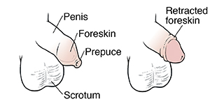
Figure 1. Tight preputial orifice on retraction of foreskin: A) Skin at preputial outlet is healthy with no scarring, and the inner preputial mucosa is starting to evert through the outlet. With physiologic phimosis, the preputial outlet is always closed and one cannot see the glans unless the foreskin is retracted, as the examiner has done in the photograph. B) In many cases of pathologic phimosis, the glans and meatus are visible without any attempt at retraction, as the scarred ring holds the preputial outlet open. There is no inner mucosal eversion through the outlet. - "Pathologic and physiologic phimosis: approach to the phimotic foreskin."
Potentially under-recognized late-stage physical and psychosexual

Single-cell analysis of human prepuce reveals dynamic changes in gene regulation and cellular communications, BMC Genomics

Retrospective analyses on preputioplasties in boys with

Topical Triamcinolone for Persistent Phimosis

Phimosis SpringerLink

Pathologic Phimosis - DoveMed

Prevalence of Phimosis in Males of All Ages: Systematic Review

PDF] Phimosis--a diagnostic dilemma?

Endothelial to Mesenchymal Transition: Role in Physiology and in
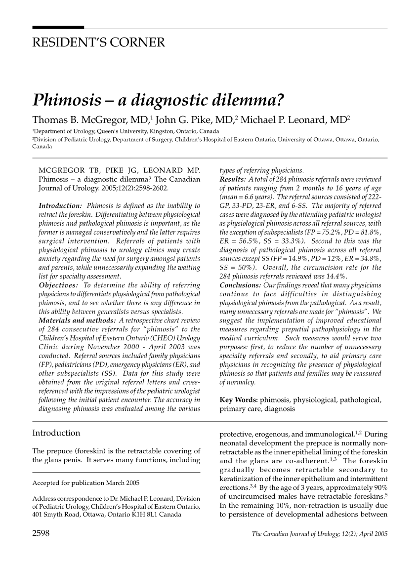
PDF) Phimosis--a diagnostic dilemma?

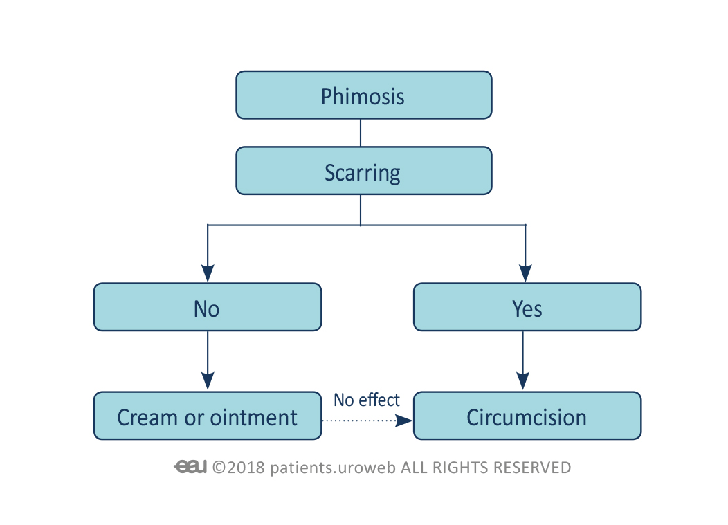
Efficacy of topical steroid therapy for phimosis treatment: a
Phimosis - Patient Information